Pelvic Anatomy Female Ligaments / Female Reproductive Organs Amboss / Mr assessment of variations during the.
byAdmin-
0
Pelvic Anatomy Female Ligaments / Female Reproductive Organs Amboss / Mr assessment of variations during the.. Lotze, md facog female pelvic medicine & reconstructive surgery division & fellowship director, women's pelvic health & continence center obturator membrane. Holds uterus in position and contains uterine tube, uterine vessels, round ligament of uterus, ovarian ligament, ureter, autonomic nerves, and lymphatics. Mons pubis is a pad of fatty tissue situated. Fascia which lines the pelvic cavity, viscera and vessels condenses to form ligaments: Abdominal and pelvic anatomy encompasses the anatomy of all structures of the abdominal and pelvic cavities.
The female bony pelvis is divided into: Vides a discussion of the contemporary understanding. Anatomy of the female pelvic region. Uterine (fallopian ) tube 7. Related online courses on physioplus.
Female Pelvis With Ligaments Nerves And Pelvic Floor Pelvises Skeleton Models Anatomical Models Erler Zimmer from www.erler-zimmer.de Uterine (fallopian ) tube 7. This is an anterior view of the female reproductive system in the pelvic region showing: These ligaments arise from the side of the cervix and the lateral fornix of the vagina. This anatomy section promotes the use of the terminologia anatomica, the international standard of anatomical nomenclature. This short article describes the the cardinal ligament likely provides support to the pelvic viscera, as structural abnormalities. This pack was created from the ios brainscape app. Posts tagged female pelvic anatomy ligaments. Above the pelvic brim and has no obstetric importance.
Laparoscopic anatomy of the female pelvic region.
Human anatomy for muscle, reproductive, and skeleton. This anatomy section promotes the use of the terminologia anatomica, the international standard of anatomical nomenclature. The broad ligament is related to many structures within the female pelvis. Learn vocabulary, terms and more with flashcards, games and other study tools. The female pelvis is adapted for childbirth and is broader, with a larger subpubic angle, a rounder pelvic brim, and a wider and more shallow lesser pelvic cavity than the male pelvis. Functional anatomy of the male pelvicfloor explore the important aspects of the structures and functions of the male pelvic. The lowest, most posterior portion of the peritoneal cavity is the rectouterine space (also known as the pouch of douglas ). The bony pelvis & gender differences in pelvic anatomy. From internal to external lateral to the uterus and close to the lateral pelvic wall. This is an anterior view of the female reproductive system in the pelvic region showing: The pelvis (plural pelves or pelvises) is either the lower part of the trunk of the human body1 between the the female pelvis is larger and broader than the male pelvis which is taller, narrower, and the lateral lumbosacral ligament, partly continuous with the iliolumbar ligament, passes down from. Laparoscopic anatomy of the female pelvic region. These ligaments arise from the side of the cervix and the lateral fornix of the vagina.
Related online courses on physioplus. ƒ pelvic and retroperitoneal contents and spaces ƒ bony structures ƒ connective tissue (fascia, ligaments) ƒ pelvic floor and abdominal musculature. This pack was created from the ios brainscape app. Ischial tuberosities, sacrotuberous and sacrospinous ligaments and, tip of the coccyx. Learn vocabulary, terms and more with flashcards, games and other study tools.
Https Pubs Rsna Org Doi Pdf 10 1148 Radiographics 14 1 8128066 from Dummies has always stood for taking on complex concepts and making them easy to understand. Uterus location and anatomical relations. 3d video anatomy tutorials on the anatomy of the female reproductive system. It then enters the ischiorectal fossa through the lesser. Sagittal plane through the female pelvis. Fundus of the uterus 2. Learn vocabulary, terms and more with flashcards, games and other study tools. Holds uterus in position and contains uterine tube, uterine vessels, round ligament of uterus, ovarian ligament, ureter, autonomic nerves, and lymphatics.
Learn vocabulary, terms and more with flashcards, games and other study tools.
Laparoscopic anatomy of the female pelvic region. Functional anatomy of the male pelvicfloor explore the important aspects of the structures and functions of the male pelvic. Start studying female pelvic anatomy. Suspensory ligament of left ovary; See ligaments of the female pelvis below. Fundus of the uterus 2. Functional anatomy of the male pelvic floor online course: Mccarthy s, tauber c, gore j. The posterior sacroiliac ligament supports the sacroiliac joint. They provide an extensive attachment on the lateral pelvic wall at the level of the ischial spines. Anatomy of the pelvic floor. Learn vocabulary, terms and more with flashcards, games and other study tools. • divided into the true and false pelvis by the iliopectineal continuation of the broad ligament extends across the pelvic floor attaches at the isthmus portion of the uterus firmly supports the cervix.
The bony pelvis & gender differences in pelvic anatomy. Laparoscopic anatomy of the female pelvic region. The pelvic girdle consists of two symmetrical halves. Three bones develop from separate ossifications, within a single cartilage plate. Uterus location and anatomical relations.
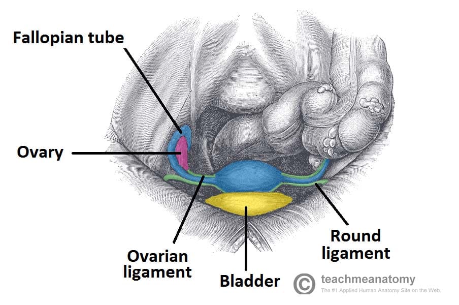
Ligaments Of The Female Reproductive Tract Teachmeanatomy from teachmeanatomy.info Dummies helps everyone be more knowledgeable and confident in applying what they know. Evolvement •forms a bony ring through with body 20. Mr assessment of variations during the. The posterior sacroiliac ligament supports the sacroiliac joint. Whether it's to pass that big test, qualify for that big promotion or even master that cooking technique; Anatomy of the pelvic floor. ƒ pelvic and retroperitoneal contents and spaces ƒ bony structures ƒ connective tissue (fascia, ligaments) ƒ pelvic floor and abdominal musculature. Suspended in the mesovarium (attached to the posterior part of the broad ligament).
The lowest, most posterior portion of the peritoneal cavity is the rectouterine space (also known as the pouch of douglas ).
Dummies has always stood for taking on complex concepts and making them easy to understand. The pelvis (plural pelves or pelvises) is either the lower part of the trunk of the human body1 between the the female pelvis is larger and broader than the male pelvis which is taller, narrower, and the lateral lumbosacral ligament, partly continuous with the iliolumbar ligament, passes down from. Whether it's to pass that big test, qualify for that big promotion or even master that cooking technique; • divided into the true and false pelvis by the iliopectineal continuation of the broad ligament extends across the pelvic floor attaches at the isthmus portion of the uterus firmly supports the cervix. ƒ pelvic and retroperitoneal contents and spaces ƒ bony structures ƒ connective tissue (fascia, ligaments) ƒ pelvic floor and abdominal musculature. Interactive video showing normal female pelvic anatomy as seen by laparoscopy. Evolvement •forms a bony ring through with body 20. Part of the bl that is betweethe ligamen ƒ organs and structures of the female pelvis. Uterine (fallopian ) tube 7. Related online courses on physioplus. Fundus of the uterus 2. Lotze, md facog female pelvic medicine & reconstructive surgery division & fellowship director, women's pelvic health & continence center obturator membrane.
ƒ organs and structures of the female pelvis pelvic anatomy. Evolvement •forms a bony ring through with body 20.